What is it?
It involves high-frequency sound waves being exposed to the body to produce images of the inside. They can show the structure and movement of organs taken in real-time. Blood flow can be looked at as well in exceptional detail. It includes 3-D pictures taken where the sound wave is converted to an image by a computer. Advanced ultrasound even allow four-dimensional capabilities (movement of 3-D).
Common Uses
Help evaluate symptoms such as pain, swelling and infection.
Examine the body internal organs such as,
- heart and blood vessels, including the abdominal aorta and its major branches
- liver
- gallbladder
- spleen
- pancreas
- kidneys
- bladder
- uterus, ovaries, and unborn child (fetus) in pregnant patients
- eyes
- thyroid and parathyroid glands
- scrotum (testicles)
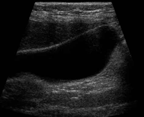
Image of the gallbladder
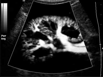
Inside the liver. The tiny blood vessels look like the branches from a tree.
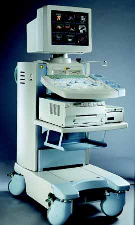
Equipment
It consists of a computer, display screen, and transducer. The transducer sends out the sound waves and then listens for returning echos from tissues in the body. It is attached by a cord to the main console.